
12 Oct 5 Ways to Use Ophthalmic Imaging Data to Educate Patients
As an eye care professional, it is important to remember that your patients have different knowledge than you do about diseases and terminology. Educating patients using ophthalmic imaging tools promotes proactive eye care and improves treatment results.
However, it can be challenging to translate complex information into understandable patient insights. Here are five effective strategies for using ophthalmic imaging data to educate your patients.
1. Implement an interactive ophthalmic imaging system that integrates with your EHR.
One problem related to using ophthalmic imaging data to educate patients is the need for more engagement and understanding among patients. Many patients need help grasping the technical aspects of their eye conditions and the significance of the imaging data you are showing them.
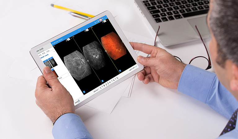
Bridge the gap between complex ophthalmic imaging data and patient understanding by implementing an interactive cloud-based ophthalmic image management system such as EyeClinic Imaging.
EyeClinic Imaging enhances patient engagement and empowers individuals to take a more active role in their personal eye care—leading to better treatment adherence and improved eye health outcomes.
The most effective ophthalmic image management systems seamlessly integrate with various ophthalmology and optometry EHR solutions, like MaximEyes.com. Alternatively, many practices use ophthalmic imaging as a standalone tool when needed.
For instance, EyeClinic Imaging integrates with any optometry and ophthalmology EHR system using HL7®, DICOM®, and non-DICOM interfaces and provides effortless one–click access directly from your EHR.
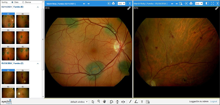
EyeClinic Imaging: Compare prior exam images side-by-side.
“Images and testing results automatically upload into EyeClinic Imaging. I can review them with a single click directly from within MaximEyes.com EHR. I love viewing images side-by-side to detect and annotate any changes. EyeClinic Imaging helps me educate patients on why they need these tests.” –Dr. Peter Falk
2. Visualize eye anatomy using a high-resolution optical imager.
One of the most fundamental uses of ophthalmic imaging data is to help patients understand the complex anatomy of their eyes.
Many patients have limited knowledge of the intricate structures within their eyes, and visualizing these structures through imaging is incredibly enlightening to many patients. Patients better understand how their eyes function by displaying high–resolution cornea, lens, retina, and optic nerve images.
For example, retinal imaging provides detailed pictures of the retina’s various layers and blood vessels. This is especially beneficial when explaining conditions like age-related macular degeneration (AMD) or diabetic retinopathy, allowing patients to see the affected areas and comprehend the importance of early detection and treatment.
Comparing before–and–after images following treatment or surgery is another great way to educate patients. This method demonstrates the effectiveness of cataract surgery, LASIK, or intravitreal injections.
Patients witness the improvements in their eye health and vision by comparing images taken before the procedure with those taken after. This visual boosts their confidence in the chosen treatment and encourages patients to follow post-treatment care instructions diligently.
3. Track eye disease progression and chronic conditions.
Ophthalmic imaging data is also used to track the progression of eye diseases over time. Imaging data is particularly valuable for chronic conditions such as glaucoma. Visually demonstrate how the disease advances or stabilizes by showing patients sequential images of their optic nerve and visual field tests.
Showing these changes visually motivates patients to adhere to treatment plans, including taking prescribed medications and attending regular check-ups. It also helps them recognize the significance of monitoring their eye health, even when they may not experience noticeable symptoms.
From visualizing eye anatomy to tracking disease progression, optical imaging provides patients valuable insights into their eye health.
Rather than spending valuable time searching for digital optical images and manually associating them with the patient’s medical record or resorting to the laborious process of printing and scanning files, you effortlessly access and review images for any patient with a single click.
Related: 5 Benefits of an Ophthalmic Image Management System
4. Illustrate and easily explain vision disorders.
With ophthalmic imaging data, explain various vision disorders to patients comprehensively and engagingly. For instance, optical coherence tomography (OCT) produces cross-sectional retina images, making it easier to illustrate conditions such as macular holes, retinal detachments, or vitreomacular traction.
Displaying these images and pointing out the affected areas simplifies complex medical terminology and helps patients grasp the nature of their condition.
Moreover, these visuals emphasize the importance of treatment options and lifestyle changes that improve or stabilize their vision. EyeClinic Imaging allows you to easily select and view images side-by-side, zoom, pan, notate, and more.
“Our patients are very impressed that we can bring up their test results fast, especially fundus camera photos while they are still in the chair. It’s like they say, ‘A picture is worth a thousand words.’” –Dr. Brandon Chester
5. Personalize treatment plans for each patient’s unique profile.
One of the most powerful ways to use ophthalmic imaging data in patient education is by tailoring treatment plans to each patient’s unique visual profile. Not all eyes are the same; optical imaging allows for a highly personalized approach to vision care.
For example, when discussing refractive surgeries like LASIK or Photorefractive Keratectomy (PRK), corneal topography maps demonstrate the specific irregularities in the patient’s cornea. Patients will better understand why you recommend a particular surgical technique and what outcomes they should expect based on their eye measurements.
In retinal diseases, such as diabetic retinopathy, fluorescein angiography reveals the extent of blood vessel damage. Sharing these images with the patient underscores the necessity of strict blood sugar control, regular eye examinations, and the potential benefits of treatments like laser therapy or anti-VEGF injections.
“I highly recommend EyeClinic Imaging to my colleagues—it’s the future of image management for the private practice. I love how easy it is to upload images. Combining all images is great clinically as it saves time and is perfect for educating the patient.” –Dr. Gil Catino
Educate, Engage, and Empower Patients Through Cloud-Based Ophthalmic Imaging
Leveraging ophthalmic imaging data that integrates with your EHR is the new norm to educate patients and foster proactive eye care. You put your patients in control as active partners in their eye care journey—making informed decisions and taking steps to protect and preserve their vision.
The patient education journey starts with having the right ophthalmic imaging system in place so you deliver more value to your patients. EyeClinic Imaging combines all your diagnostic imaging so you can easily access your data to educate, engage, and empower your patients.